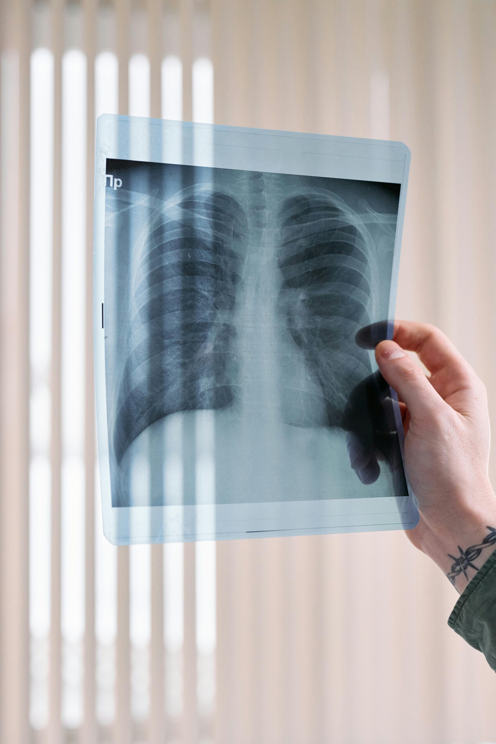
Chest X-RAYS
Discover what your chest X-ray reveals — a simple breakdown of what’s hiding in plain sight.
Chest X-Rays: What They Are and How They Work
Chest X-rays are one of the most commonly performed imaging tests in medicine, offering a quick and reliable way to assess the health of your heart, lungs, and other structures inside your chest. But have you ever wondered how they actually work or what doctors are looking for when interpreting them?
Whether you’ve had a chest X-ray yourself or are simply curious about this fascinating tool, understanding its basics can give you a clearer idea of its role in healthcare.
In this article, we’ll explore what a chest X-ray is, how it works, and how doctors interpret it — all in simple terms. Let’s dive in!
What Is a Chest X-Ray?
A chest X-ray (CXR) is a type of medical imaging test that uses a small amount of radiation to create pictures of the inside of your chest. These images help doctors look at your lungs, heart, blood vessels, and even your ribs to check for any abnormalities or health conditions.
X-rays work because they can pass through your body but interact differently with tissues depending on their density. For example:
- Air (like in your lungs) appears dark or black because it doesn’t block much of the X-ray beam.
- Bone (like your ribs) looks white because it blocks most of the X-rays.
- Soft tissues (like your heart) appear in shades of gray, somewhere in between.
This contrast helps doctors pinpoint issues, such as fluid buildup, infections, or other abnormalities.
How Does It Work?
During a chest X-ray, a machine emits a controlled beam of X-rays that pass through your chest and hit a detector (either film or digital). The resulting image is a snapshot of how different structures in your chest interact with the X-ray beam.
There are two common views of a chest X-ray:
1. Posteroanterior (PA): The X-ray beam passes from your back to your front. This view is ideal for accurately assessing the size of your heart.
2. Anteroposterior (AP): The X-ray beam passes from your front to your back. This view is often used in hospitalized patients who can’t stand but can make the heart appear slightly larger.
Understanding these views is important for accurate interpretation of the images.
What Does a Normal Chest X-Ray Look Like?
Before identifying a problem, it’s essential to know what “normal” looks like. On a healthy chest X-ray, you’ll see:
- Lungs that appear clear with no unusual shadows or densities.
- A heart that is appropriately sized (usually less than half the width of the chest in a PA view).
- Bones, like ribs and the spine, without fractures or deformities.


How Do Doctors Identify Problems on a Chest X-Ray?
When something is wrong, it often shows up in specific patterns:
- Pneumothorax (Collapsed Lung): A sharp white line marks where the lung has collapsed, and the surrounding area may look unusually dark.
- Pulmonary Edema (Fluid in the Lungs): White spots or haziness appear in the lungs due to fluid buildup.
- Pleural Effusion (Fluid Around the Lungs): A curved white line (known as a meniscus) at the bottom of the lung cavity indicates fluid collection.
Each finding helps guide doctors in diagnosing and treating the underlying condition.
Why Are Chest X-Rays Important?
Chest X-rays are a vital tool in medicine. They’re quick, non-invasive, and can provide a wealth of information about your health. From spotting infections to confirming the placement of medical devices like breathing tubes, they play an essential role in diagnosing and monitoring many conditions.
Understanding how chest X-rays work and what they show can give you valuable insight into your health and help you feel more confident during your next visit to the doctor.
In summary, a chest X-ray is more than just a picture — it’s a window into your body’s inner workings!Chest X-Rays: What They Are and How They Work
Chest X-rays are one of the most commonly performed imaging tests in medicine, offering a quick and reliable way to assess the health of your heart, lungs, and other structures inside your chest. But have you ever wondered how they actually work or what doctors are looking for when interpreting them?
Whether you’ve had a chest X-ray yourself or are simply curious about this fascinating tool, understanding its basics can give you a clearer idea of its role in healthcare.
In this article, we’ll explore what a chest X-ray is, how it works, and how doctors interpret it — all in simple terms. Let’s dive in!
What Is a Chest X-Ray?
A chest X-ray (CXR) is a type of medical imaging test that uses a small amount of radiation to create pictures of the inside of your chest. These images help doctors look at your lungs, heart, blood vessels, and even your ribs to check for any abnormalities or health conditions.
X-rays work because they can pass through your body but interact differently with tissues depending on their density. For example:
- Air (like in your lungs) appears dark or black because it doesn’t block much of the X-ray beam.
- Bone (like your ribs) looks white because it blocks most of the X-rays.
- Soft tissues (like your heart) appear in shades of gray, somewhere in between.
This contrast helps doctors pinpoint issues, such as fluid buildup, infections, or other abnormalities.
How Does It Work?
During a chest X-ray, a machine emits a controlled beam of X-rays that pass through your chest and hit a detector (either film or digital). The resulting image is a snapshot of how different structures in your chest interact with the X-ray beam.
There are two common views of a chest X-ray:
- Posteroanterior (PA): The X-ray beam passes from your back to your front. This view is ideal for accurately assessing the size of your heart.
- Anteroposterior (AP): The X-ray beam passes from your front to your back. This view is often used in hospitalized patients who can’t stand but can make the heart appear slightly larger.
Understanding these views is important for accurate interpretation of the images.
What Does a Normal Chest X-Ray Look Like?
Before identifying a problem, it’s essential to know what “normal” looks like. On a healthy chest X-ray, you’ll see:
- Lungs that appear clear with no unusual shadows or densities.
- A heart that is appropriately sized (usually less than half the width of the chest in a PA view).
- Bones, like ribs and the spine, without fractures or deformities.
How Do Doctors Identify Problems on a Chest X-Ray?
When something is wrong, it often shows up in specific patterns:
- Pneumothorax (Collapsed Lung): A sharp white line marks where the lung has collapsed, and the surrounding area may look unusually dark.
- Pulmonary Edema (Fluid in the Lungs): White spots or haziness appear in the lungs due to fluid buildup.
- Pleural Effusion (Fluid Around the Lungs): A curved white line (known as a meniscus) at the bottom of the lung cavity indicates fluid collection.
Each finding helps guide doctors in diagnosing and treating the underlying condition.
Why Are Chest X-Rays Important?
Chest X-rays are a vital tool in medicine. They’re quick, non-invasive, and can provide a wealth of information about your health. From spotting infections to confirming the placement of medical devices like breathing tubes, they play an essential role in diagnosing and monitoring many conditions.
Understanding how chest X-rays work and what they show can give you valuable insight into your health and help you feel more confident during your next visit to the doctor.
In summary, a chest X-ray is more than just a picture — it’s a window into your body’s inner workings!

